
-
-
-
-
Service

-
Contact Us

Product Details
product information
【product name】
Product Name: Fungal Double Fluorescent Staining Solution
Common Name: Fungal Double Fluorescent Staining Solution
【Packaging Specifications】
Product name: Type I
Specifications: □100 servings/bottle 2ml □200 servings/bottle 4ml
【Period of use】
1. Store at room temperature and in the dark, do not freeze, valid for 3 years from the date of production.
2. Use directly after opening the bottle, and it can be stable for 180 days at room temperature after opening.
【Operating procedures】
1. Skin flakes, nail flakes, and hair samples are scraped and placed on a glass slide, drop a drop of staining solution, cover with a cover glass, and observe under a fluorescence microscope.
2. For body fluids and vaginal secretions, take a small amount and apply it to a glass slide, add a drop of staining solution, cover it with a cover glass, and observe it under a fluorescence microscope.
3. To determine whether there is fungi in the sample, it can be completed under the microscope of 100 times and 400 times; when the bacteria identification and CYP51 protein expression are judged according to the double fluorescent staining solution, it needs to be observed under the oil microscope. Please refer to the relevant map for bacterial identification methods.
【Basic Information】
Product record number: Yuesui Machinery No. 20181119
Medical device production record certificate number: Yuesui Food and Drug Supervision Equipment Production No. 20180177
【Storage conditions】
Store at room temperature and dark, do not freeze.
【Instrument requirements】
Microscope with 340-380nm filter.
【Precautions】
1. This kit is only for in vitro detection and diagnosis, please read the instructions carefully before use.
2. This kit is limited to the qualitative analysis of fungi.
3. This kit needs to be stored at room temperature away from light, and must not be frozen.
4. The reagent is corrosive to a certain extent, please handle it with caution when using it. Do not swallow.
5. During operation, avoid direct sunlight or strong light.
6. When discarding reagents and disposal, please follow the relevant regulations on waste.
7. Reagents of different batches cannot be mixed.
Fungal Double Fluorescent Fluorescent Staining Solution
Product principle
The fungal cell wall contains chitin, and the interior of the cell contains CYP51 protein (n-lanosterol 14a demethylase P450-14DM CYP51). The main components of this product are fluorescent whitening agent and CYP51 protein fluorescent dye.
(1) The fungal cell wall chitin can be stained by fluorescent whitening agents.
(2) The distribution of CYP51 protein in different species of fungi is different.
(3) Since CYP51 protein is an intracellular protein, when the fungal activity is good, the amount of CYP51 protein fluorescent dye into the fungal cells is small.
(4) The expression of CYP51 protein can cause fungi resistance to azoles. Therefore, bacterial species identification or fungal activity identification can be carried out according to the above characteristics.
The above principles are applicable .
Introduction
Detect various superficial and deep fungal disease specimens. For tinea corporis, tinea pedis, onychomycosis, tinea capitis, tinea versicolor, deep fungal infections , etc., fungal inspection, determination of bacterial cell death and survival, identification of bacterial species, and preliminary determination of azole resistance were carried out.
Feature 1
Judging the fungus dead and alive, it is used for curative effect detection, avoiding over-medication . The hyphae and spores of the surviving fungi are bright blue on the outside, pink , and the nucleus is clearly visible. Dead fungal hyphae and spores are bright blue on the outside and dark red on the inside, and nuclei are sometimes visible.
Feature 2
Display subcellular structure, provide rich information for bacterial species identification. In addition to displaying fungal cell walls and hyphal intervals, this product can clearly display the size and number of nuclei, endoplasmic reticulum and other subcellular structures. Different fungi have different nuclei and endoplasmic reticulum, so the fungal species can be judged by double fluorescent staining. This product can display the sub-new cell structure of skin fungi, Malassezia, Candida, Sporothrix, color fungi, and molds .
Feature three
The CYP51 protein content was shown to assist in the identification of azole drug susceptibility to CYP51-overexpressing fungi, and the bacteria were obviously clump-shaped, suggesting that the fungi were resistant to azole drugs. Non-drug-resistant bacteria have no clump-like structure inside. This product can preliminarily suggest the sensitivity of fungi to commonly used azole drugs such as itraconazole and ketoconazole. In case of drug-resistant fungi, Echinococcus can be used instead of pharmacological antifungal drugs.
Fungal Microscopy Reporting System
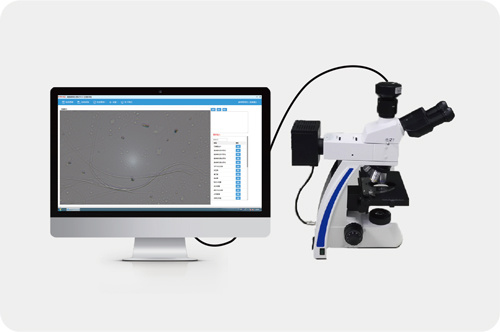
Software Features
◈ Humanized man-machine interface
◈ The software is concise, easy to operate, easy to understand and easy to maintain
◈ Data is safe and reliable, data can be backed up and exported
Features
◈ Obtain clear image information of fungal mirror, and realize the synchronous display of image information on the workstation side.
◈ Perform image collection to form a graphic diagnosis report.
◈ High-definition large-screen display of details under the microscope, clear imaging, and synchronization with the fungal microscope.
◈ Realize the management, analysis, statistics and other functions of patient data and image data information
Key words:
fungal
fungi


Company address: Guangzhou, No. 3, Lanyue Road, Huangpu District, Guangzhou International Business Incubator F113
Fax:020-87393579

scan it
Follow Chuanghong Dynamics
Copyright © 2018 Guangzhou Chuanghong Medical Technology Co., Ltd. 粤ICP备14098993号 SEO
Tel: 400-688-0628
Company address: F113, Guangzhou International Business Incubator, No. 3 Lanyue Road, Huangpu District, Guangzhou
E-mail: chuanghongmedical@gmail.com
We will give you feedback in time